


Modern visual restoration operations are high -tech and safe procedures that allow us to eliminate almost all ophthalmic problems.They have been successfully used for several decades, so the methods develop, expand and become constantly more effective.The improvement of visual functions is obtained using the hardware correction of the shape of the cornea, the lenses, the retina and other elements of the optical eye system.The correctly selected technology allows not only to completely restore the vision, but also to reduce the risk of complications.From the article you will learn what there are ophthalmic operations, indications for use and possible risks.
Types
Thanks to the development of medical hardware methods, the visual and minimally invasive procedures are reliable and minimally invasive.Their duration does not exceed several hours and in the future complex rehabilitation measures are needed.The choice of the surgical treatment method is chosen on the basis of the disease, age and general conditions of the patient's visual apparatus.
Laser correction
The most popular type of operation to correct visual acuity.Today these are high -efficiency high -technology methods and a minimum risk of complications.Allow you to cope with myopia, foresight and abstigmatism.After the procedure, the visual acuity is maintained for a long time and if you follow all the instructions of an ophthalmologist, it is possible to completely avoid repeated intervention.There are different types of laser correction:
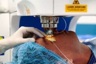
- Lasik.The type of basic operation to restore visual acuity.First of all, the superficial layer of the cornea is separated from a microcate and therefore, using a laser beam, a change is performed in its shape.The main disadvantage of this type of correction is the inability to take into account the individual characteristics of the anatomy of the patient's eyes;
- Super Lasik.An advanced version of the traditional Lasik methodology.It allows you to obtain a better result, as it takes into account the structure of the patient's visual system.Used in most modern clinics in the world;
- Femto Lasik.A type of similar operation, the only difference is that the cutting of the cornea is performed not by a microcarat, but by a special Femo laser.There is an improved version in which the course of the operation depends on the patient's individual characteristics - Super Femto Lasik;
- Epi-Lasik.The mechanism of the procedure is identical to the traditional Lasik method, but this operation is prescribed only to patients with thin horny (acquired or congenital);
- PRK (FRK).The photographic refraction cheratectomy has been performed since 1985. Today it is applied in the presence of contraindications to ordinary correction methods, for example with a thin cornea, serious ophthalmic diseases.The healing process is always painful, the recovery period lasts longer than other methods.
Visual correction operations do not last more than 15 minutes.After the procedure, it is necessary to wear a protective dressing for several hours, as well as the instillation of drops for 1-2 months.The risk of complications is minimal, repeated treatment is needed with a significant reduction in vision.
Vitreltomy
This is a procedure for the complete or partial removal of the vitreous body of the eyeball.It is carried out under general or local anesthesia, in the absence of complications, passes in 2-3 hours.First of all, small drips are carried out in the eye socket, through which subsequent manipulations are performed.As a rule, this is a cauterization by a laser of the affected areas of the retina, the densification of abroad or the restoration of the integrity of the fabric.The procedure is prescribed for the following problems:
- Restoration of visual functions after hemorrhage in the fabrics of the eye;
- Prevention of the detachment of the retina related to age;
- The treatment of the serious retinopathy of the eyes in which gross or neovascularization scars occur (germination of the blood vessels).
Artificial polymers, gas bubble, silicone oil or a balanced salt solution are used to replace the vitreous body.The latter type is used more often, since in the future a repeated operation is not required: the saline solution is subsequently replaced by the intraocular fluid.
After surgery, side effects are possible in the form of swelling of the cornea, increased intraocular pressure or even more vision.The restoration and forecasts depend on the vastness of the lesion, as well as the type of prostheses when replacing the vitreous body.If there are irreversible changes in the optical nerve, the correction of vision is almost impossible.
Scleroplasty
A common ophthalmic procedure aimed at strengthening the external shell of the eye (sclera).It is prescribed not to correct visual functions, but to stabilize the degree of myopia in the patient from the risk group.It is recommended to be carried out by teenagers who suffer from this problem, since at this age the shape of the eye is actively changing.
During the operation, the required quantity of flaps of material to strengthen the sclera is introduced behind the rear wall of the eyeball.Biological polymers or components are generally used.Subsequently, a peak occurs with the external shell of the eye and after a few months the blood vessels needed to maintain visual functions grow in the flap.There is a simplified version of scleroplasty.It is an introduction of an artificial or biological substance for the eyeball.The mechanism of action in this technology is identical, preventing the growth of the eyeball.
Scleroplasty
This is a well -studied operation that practically does not change over the years.It is performed in most clinics.There have been practically no side effects, with the exception of possible drug allergies.A second operation is usually required.
Replacement of the objective
The necessary operation, which is prescribed during clouding or any other degenerative process in the lens, for example cataracts.The treatment is always forced, but the system is selected individually, depending on the age, sex and severity of pathological changes in the eye.The replacement of the crystal is prescribed in the following cases:
- High degrees of myopia and foresight;
- a significant reduction of refraction;
- regenerative processes in the eye, reduction of the vision related to age;
- the impossibility of restoring laser vision;
- cataract;
- The probability of developing glaucoma on a background of a systemic or ophthalmic disease.
The procedure always takes place under local anesthesia.During the operation, the surgeon makes a small incision with a laser, after which the patient's goal dilutes with a special tool and removes it from the eye.Subsequently, a prepared transplant is installed.The intervention does not last more than 25 minutes, the subsequent imposition of seams and recovery in the hospital is not required.
The operation is performed in most private and state clinics.Usually there are no complications after manipulation, but the subsequent correction of the laser vision is often prescribed.In rare cases, a reduction in the lens is required.
Keratoplasty (replacement of the cornea)
One of the most modern and complex ophthalmic operations, which is associated with many risks and requires a high qualification of the surgeon.It is necessary to restore anatomical integrity and physiological functions of the cornea.It is prescribed for the treatment of congenital or acquired defects obtained following injuries or diseases.The healthy tissue for transplantation is assumed only in donors, but the development of artificial replacement is carried out in many countries.We recommend keratoplasty to solve the following problems:
- Treatment of corneal diseases (Belko, tone disorders);
- mechanical or chemical damage;
- Congenital defects.
The operation is performed no more than 30 minutes.During the procedure, the surgeon with a laser or a special scalpel removes part of the patient's cornea and in its place plants the donor fabric.The seams can persist up to a year, after which a special lens is selected to reduce the risk of infections.The recovery period is 4 weeks, during which the antibiotic is needed, but regular exams are needed for the entire following year.
In recent years, it has been possible to significantly reduce the risk of refusal of the donor fabric due to the use of special compositions during its processing and conservation.
Laser coagulation of the retina
The operating method of restoring retinal fabric.The effectiveness of the method is greater than 70%and already 24 hours after its implementation, you can return to the usual lifestyle.The observations of an ophthalmologist are needed within one year from the procedure.
Today, the operation is performed using a laser, which allows you to do without the blood loss.Anesthesia is carried out locally, the procedure time does not require more than 20 minutes.
Before exposure to the laser, drops of drops to expand the pupil and then wear a special protective lens, through which exposure to low frequencies occurs.Due to high temperatures, glue damaged cells and small blood vessels.
The coagulation procedure is necessary for any damage and pathologies of the retina, as well as with the eyes and diseases of the vascular system of this organ.After the operation, inflammation and annex is possible.For several years after the correction, it is impossible to engage in heavy physical work and active sports.
Reticulation
An effective method for the treatment of various horny diseases.It is carried out to strengthen the ligaments and other fibers in the corneal tissues, which is necessary for the keratoconus of various degrees or degenerative processes, dystrophy.
The operation is performed under local anesthesia.First of all, a small part of the cornea is cut with a special device and vitamin B2 is instilled in the open area.The subsequent irradiation allows you to compact fabrics of over 200%.The first week after the operation, it is necessary to wear a protective contact lens and for 6 months it must be examined by a doctor.The effect of the procedure persists for 10 years, therefore a repeated operation is required.
Complications are observed in rare cases.The patient can reduce the vision, inflammation or exit of the cornea.
Glaucoma treatment
The ophthalmological operation for various degrees of glaucoma is necessary when drug therapy does not bring the necessary result.The operation is performed using a laser or surgically.
The laser method is considered the most successful.It is completely painless for the patient and practically there are no complications.During the procedure, a hole is made with a radius through which the liquid is removed from the fabrics of the eye to normalize the pressure.It is used to treat all types of glaucoma.
The type of manual surgical operation is less effective because there is a risk of complications after the procedure.As a rule, this is a deep scletectomy not practiced.The purpose of the method is to reduce intraocular pressure using a slight thinning of the corneal layer.
The effect after both types of operations is reduced over time.On average, after 5-7 years, a second operation is required.This period can be extended using competent pharmacological treatment.
Conclusions
Today, in most modern ophthalmic clinics, a series of surgical procedures are performed for vision correction.These are accurate high -tech methods with which it is possible to solve almost all eyes of the eyes.The choice of the method depends on a series of factors: age, disease, individual characteristics of the patient's visual system structure.After the operation, the effect occurs almost immediately and if all the doctor's prescriptions are observed, visual acuity can be preserved for a long time.